ICG Lymphatic Imaging

What is ICG Imaging?
ICG Lymphatic Imaging is the best way to assess lymphatic function and diagnose lymphoedema. It is regarded as the 'Gold Standard' for lymphoedema imaging.
Advancements in technology have made it possible for us to visualise the lymphatics in real-time. Using a special camera, and an injection of fluorescent medical dye, we can see the lymphatic vessels and lymph fluid in the the tissue.
This allows us to identify how they are working, where they are draining to and any lymphatic failure.
This particular method of imaging the lymphatics has also been referred to as:
-
Near Infrared Fluorescence Lymphatic Imaging (NIRFLI)
-
NIRF Imaging
-
Lymphofluroscopy Imaging
-
ICG Fluoroscopy
You could hear a combination of these key words depending on who you talk to, but the most generally accepted term now is "ICG Lymphatic Imaging".
Why ICG Imaging?
Being able to 'map' the lymphatics in real-time allows us to:
-
Offer specialist diagnosis and assessment
-
Map individual pathways
-
Allow for optimised FG-MLD
-
Improve long term outcomes and quality of living
-
Create a bespoke mapping plan utilising your unique drainage pathways
-
Tailor a management or reduction plan around your needs and lifestyle
-
Identification of early lymphatic failure
-
Identify changes and anticipate developments
-
Teach you self-management techniques you can use for life

ICG Imaging at LymphVision
Our ICG Lymphography clinics use protocols which were established by Professor JP Belgrado at the Brussels Lymphology Research Unit. Our procedure allows us to assess your full lymphatic status by the visualising and 'mapping' of your unique lymphatic pathways. This allows us unique insight when creating you a tailored treatment plan.
By combining the 'mapped' lymphatics with the techniques of Fluroscopy Guided Manual Lymphatic Drainage (FG-MLD®) we create a bespoke MLD plan which you can carry out at home as Self Lymphatic Drainage (SLD). Our patients report seeing faster results and improved outcomes.
More than just imaging...
Although you come to see us for lymphatic imaging, our clinics are about much more than that. We don't simply assess the quality or stage of your lymphatics in a brief appointment. We work to understand your lymphatics and their flow and exact pathways, and you leave knowing more about your lymphoedema than you probably thought was possible.
We can talk you through all of the treatment options which may be available, including the newest research and current innovations. We can request the perfect combination of garments or wraps for the best results, and discuss all of the unique factors affecting your personal presentation. We can provide handy advice to maximising your self treatments, and even some tips and tools you may not have heard before.
Mostly, we aim to educate you so that you have the tools to be empowered in your own care.


Is ICG Imaging Safe?
ICG Lymphatic Imaging is considered highly safe and has been used safely and successfully for over 50 years. There are minimal instances of allergic reactions reported, particularly with the low-dose subcutaneous injections employed for the procedure. The likelihood of allergic reactions is extremely rare.
Unlike some other imaging techniques, such as lymphoscintigraphy, ICG Imaging does not involve exposure to ionizing radiation. This is an advantage in terms of safety.
While there is a minimal risk of infection associated with injecting the affected arm or leg in lymphoedema cases, this risk is minimized by the use of very tiny needles and through the routine use of sterile equipment, antiseptic skin preparation and thorough infection control protocols.
The injected dye, which has a green color, may result in a temporary green staining on the skin, lasting for a few days post-test. Following ICG Imaging, individuals can promptly resume all normal activities, including exercising and driving themselves home.
ICG Lymphatic Imaging Gallery
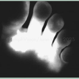
These images demonstrate the types of presentations seen during an ICG Imaging Procedure. You will be able to see your lymphatics on our high-definition screen during your procedure and our clinicans will explain the findings to you.
Sources
Figueroa, B.A.; Lammers, J.D.; Al-Malak, M.; Pandey, S.; Chen, W.F. Lymphoscintigraphy versus Indocyanine Green Lymphography—Which Should Be the Gold Standard for Lymphedema Imaging? Lymphatics 2023, 1, 25-33. https://doi.org/10.3390/lymphatics1010004


